

Room Four
Quain Vascular Anatomical Illustration 19thc - Male Pelvic Anatomy
Quain Vascular Anatomical Illustration 19thc - Male Pelvic Anatomy
Couldn't load pickup availability
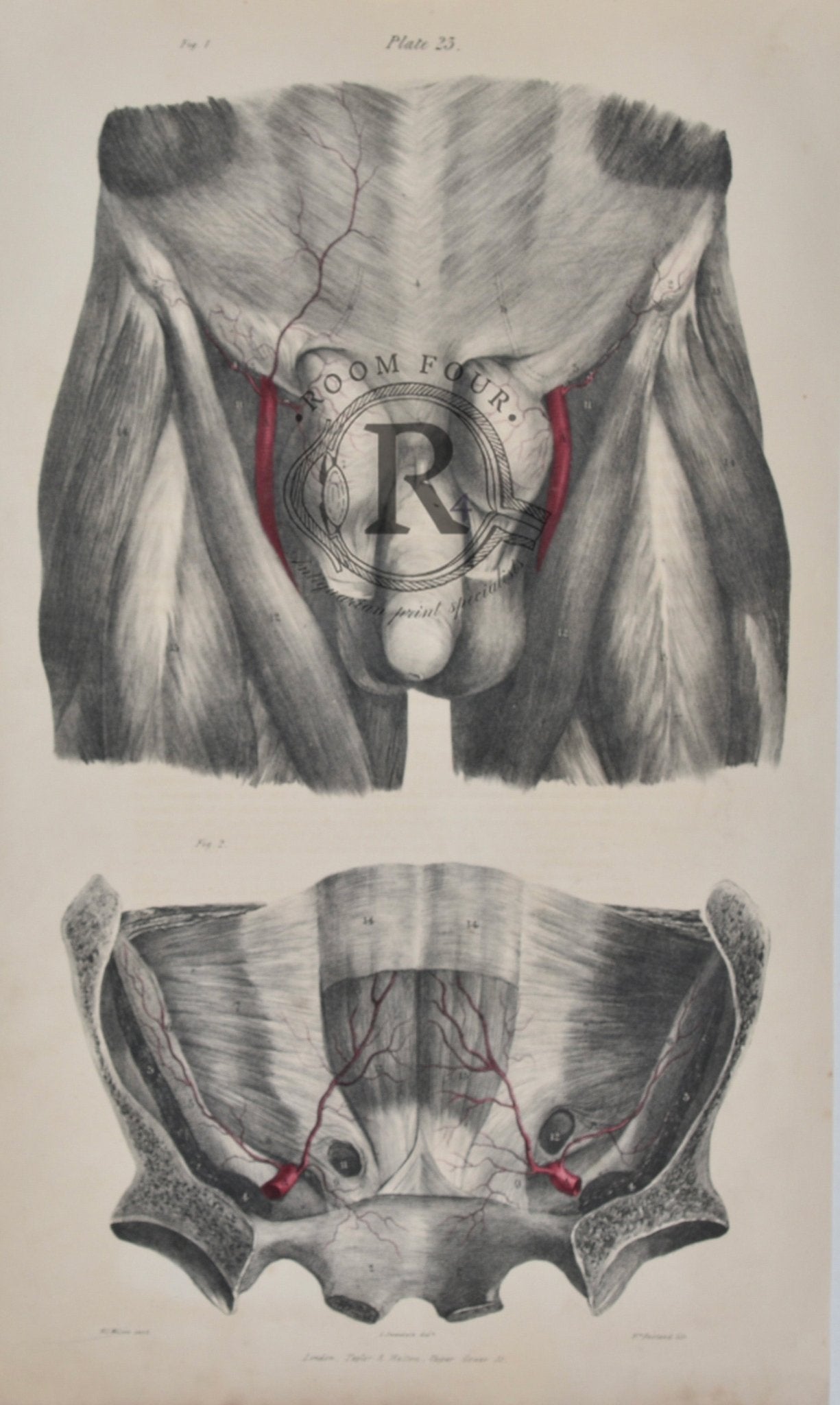
This authentic 19th‑century Quain anatomical engraving presents two detailed studies of the male pelvic region, illustrating both the superficial anatomy and the deeper vascular structures with exceptional Victorian precision. Taken from Quain’s Elements of Anatomy, one of the era’s most respected medical texts, Plate 23 captures the complexity of the pelvis through two meticulously rendered figures.
Fig. 1 depicts the external pelvic region, including the lower abdominal wall, inguinal area, proximal thigh musculature, and the superficial vessels of the scrotum and testes. The illustration highlights the femoral artery, superficial epigastric vessels, and surrounding musculature, offering a clear view of the anatomical relationships at the surface.
Fig. 2 presents a deeper dissection of the pelvic cavity, revealing the internal iliac arteries, obturator vessels, pelvic musculature, and the bony landmarks of the innominate bones. The vascular pathways are shown with remarkable clarity, demonstrating the branching patterns and connections essential to classical anatomical study.
Printed in London by Taylor & Walton, this engraving exemplifies the fine linework and scientific accuracy that make Quain’s plates highly collectible. Ideal for medical professionals, anatomy students, interior designers, and collectors of antique scientific art, this plate brings historical depth and anatomical insight to any curated space.
📏 Features & Details
-
Original 19th‑century copperplate engraving
-
Plate 23 – Pelvic anatomy (superficial & deep vascular structures)
-
Two detailed anatomical figures
-
Crisp Victorian linework and labeled structures
-
Printed by Taylor & Walton, London
-
Perfect for framing, décor, and scientific collections
🧩 Condition Statement
This antique print is in good vintage condition, with light age toning and minor edge wear typical of 19th‑century rag‑based wove paper. The image remains clear, detailed, and well‑preserved.
Original Anatomy Plate 23/RF079
Printed in London for Taylor & Walton, Booksellers and Publishers to the University of London
Circa. 1844
This original colour lithograph (now approaching 200 years old ) was used as a teaching aid for medical students and may consequently show signs of use and aging. These may include minor toning, foxing, minor tears to extremities etc
Approx. dimensions 488mm x 310mm
High Resolution Digital Format Available