Room Four
Antique Print Post Mortem Human Muscles, Organs & Vessels (Plate VII, 1800s Sibson Medical Atlas)
Antique Print Post Mortem Human Muscles, Organs & Vessels (Plate VII, 1800s Sibson Medical Atlas)
Couldn't load pickup availability
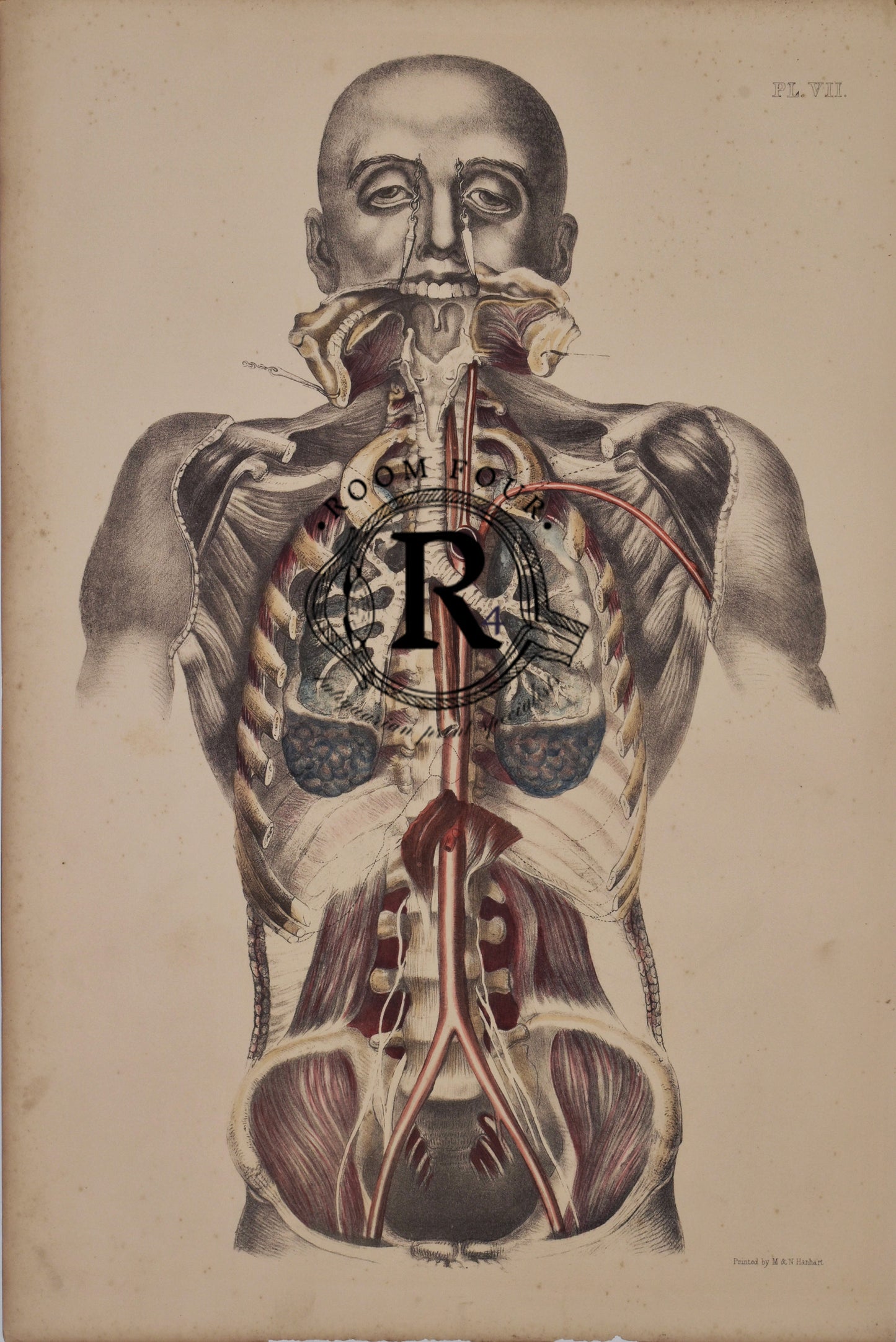
Explore the complexity of the human body with this vintage anatomical illustration featuring the internal organs and vascular system in vivid detail. Highlighting the heart, lungs, liver, stomach, kidneys, and major blood vessels, this 19th-century medical print offers both scientific accuracy and artistic elegance. Perfect for anatomy enthusiasts, educators, and collectors of antique medical art, it adds intellectual depth to gallery walls, study spaces, or cabinets of curiosities.
19th Century - Vintage Anatomical Illustration of Human Torso – Muscles, Organs & Vessels (Plate VII, 1800s Medical Atlas)
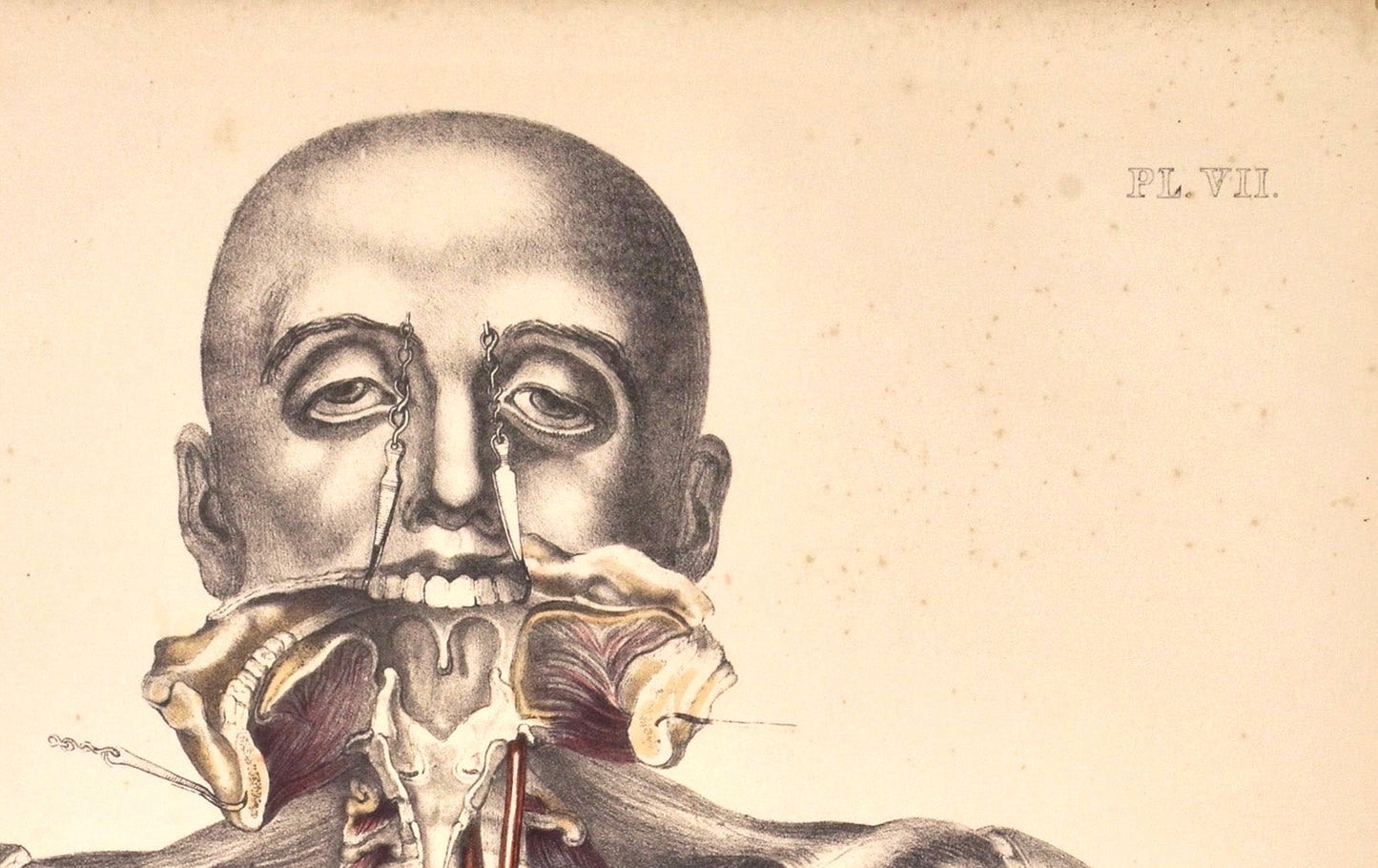
Anatomical Folio Print by Sibson – Coloured Surgical Post-Mortem Illustration - Structures of the Human Body - Muscles/Bones/Organs. This anatomical illustration—labeled PL. VII—is a classic example of 19th-century medical artistry, likely drawn for an anatomical atlas used in surgical or medical education.
Perspective: Frontal view of a male torso with the skin removed.
• Systems Shown:• Muscular system: Detailed rendering of pectoral, abdominal, and intercostal muscles, showing fiber orientation and attachment points.
• Skeletal system: Rib cage and sternum visible beneath the musculature.
• Cardiovascular system: Arteries in red, veins in blue—highlighting major vessels like the aorta and vena cava.
• Respiratory system: Lungs and trachea are exposed, with the heart nestled between.
• Digestive system: Portions of the stomach and intestines peek through the lower thoracic and upper abdominal cavity.
Artistic and Scientific Value
• Style: Meticulous linework and shading typical of copperplate engravings or lithographs from the 1800s.
• Purpose: These plates were used to teach anatomy before photography—each line drawn with surgical precision to help students visualize internal structures.
A striking and historically significant original antique folio print from the 19th century, attributed to Francis Sibson, a renowned anatomist and medical illustrator. This rare, coloured anatomical illustration features detailed surgical and post-mortem drawings, showcasing the precision and artistry of early medical documentation.
Printed during a pivotal era of scientific and medical advancement, this large-format piece was originally used as a teaching aid in medical institutions. The vivid hand-colouring and technical accuracy make it not only a valuable educational resource but also a compelling work of art.
52cm x 34cm
Key Features:
-
Authentic 19th-century print by Francis Sibson
-
Hand-coloured anatomical and surgical illustrations
-
Originally used for medical teaching and scientific study
-
Rare folio format – ideal for framing or collection
-
A unique addition to any collection of medical history, vintage science, or anatomical art
- Partially hand coloured lithographic print
This piece holds exceptional value for collectors, educators, and anyone with an appreciation for medical history, anatomical study, or antique scientific illustration.
Share