Room Four
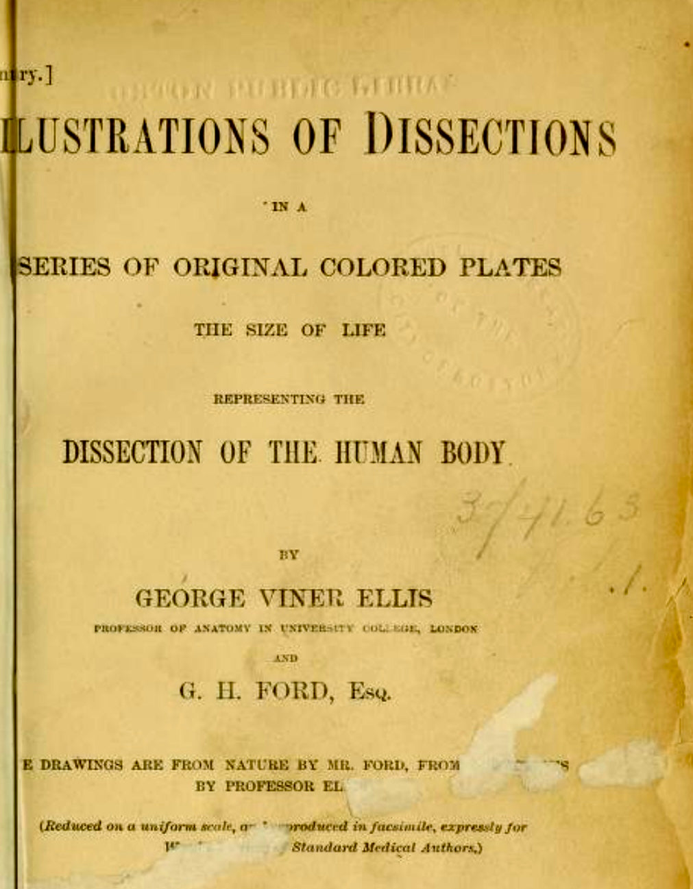
Ellis & Ford Anatomical Lithograph — Illustrations of Dissections, Plate VI — Arteries of the Arm & Shoulder (1863) | Original 19th‑Century
Ellis & Ford Anatomical Lithograph — Illustrations of Dissections, Plate VI — Arteries of the Arm & Shoulder (1863) | Original 19th‑Century
Couldn't load pickup availability
An original 1863 chromolithograph illustrating the triceps extensor muscle, shoulder musculature, arterial anatomy of the upper limb, and a clinical depiction of an olecranon fracture.
Created by George Henry Ford (1809–1876), one of the 19th century’s most respected medical illustrators, this plate was drawn life‑size from an actual dissection and printed in naturalistic colour to replicate true surgical observation.
A rare surviving teaching plate from the Victorian era, it displays the characteristic gentle toning, foxing, and light handling marks expected of an authentic 170‑year‑old anatomical print used in medical training. The detail is exceptional — Ford’s work is prized for its accuracy, clarity, and its fusion of scientific precision with artistic mastery.
A museum‑worthy collectible for medical historians, anatomy enthusiasts, physiotherapists, surgeons, or collectors of early scientific illustration.
• Artist: George Henry Ford (1809–1876)
• Date: 1863
• Technique: Chromolithograph, printed life‑size from dissection
• Subject: Triceps muscle, shoulder muscles, arterial structures, olecranon fracture
• Plate Reference: Shoulder Dissection
• Dimensions: 55cm × 37cm
• Condition: Gentle toning, foxing, and light marks consistent with age
• SKU: 004.3/26
Share