Room Four
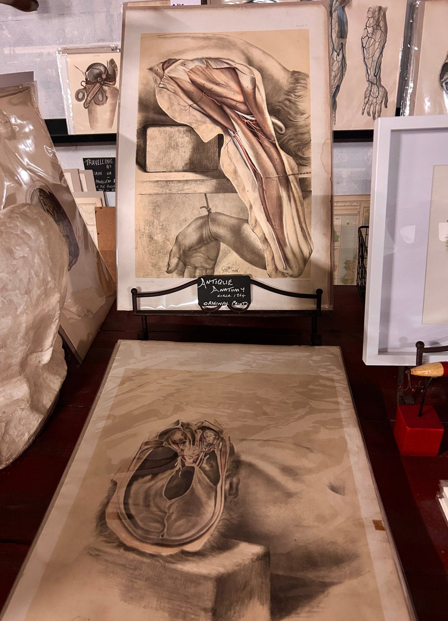
John Lizars - 19th Century Anatomical Engraving
John Lizars - 19th Century Anatomical Engraving
Couldn't load pickup availability
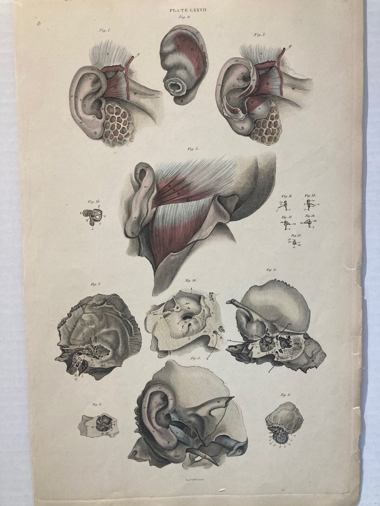
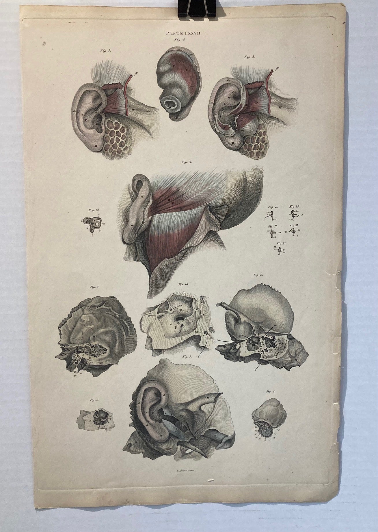
An original antique anatomical illustration of the human ear, showcasing both external and internal structures in precise detail -
Dimensions: 44cm x 28cm
Provenance:
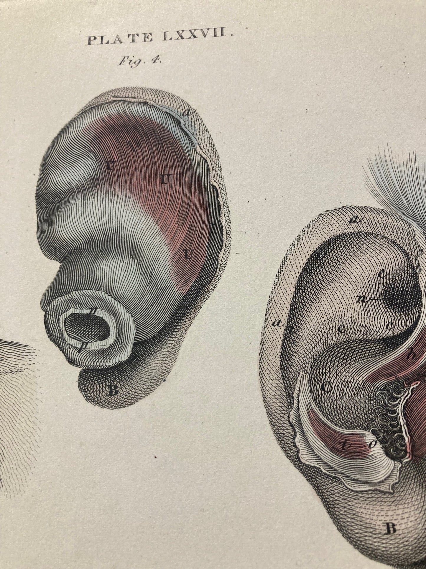
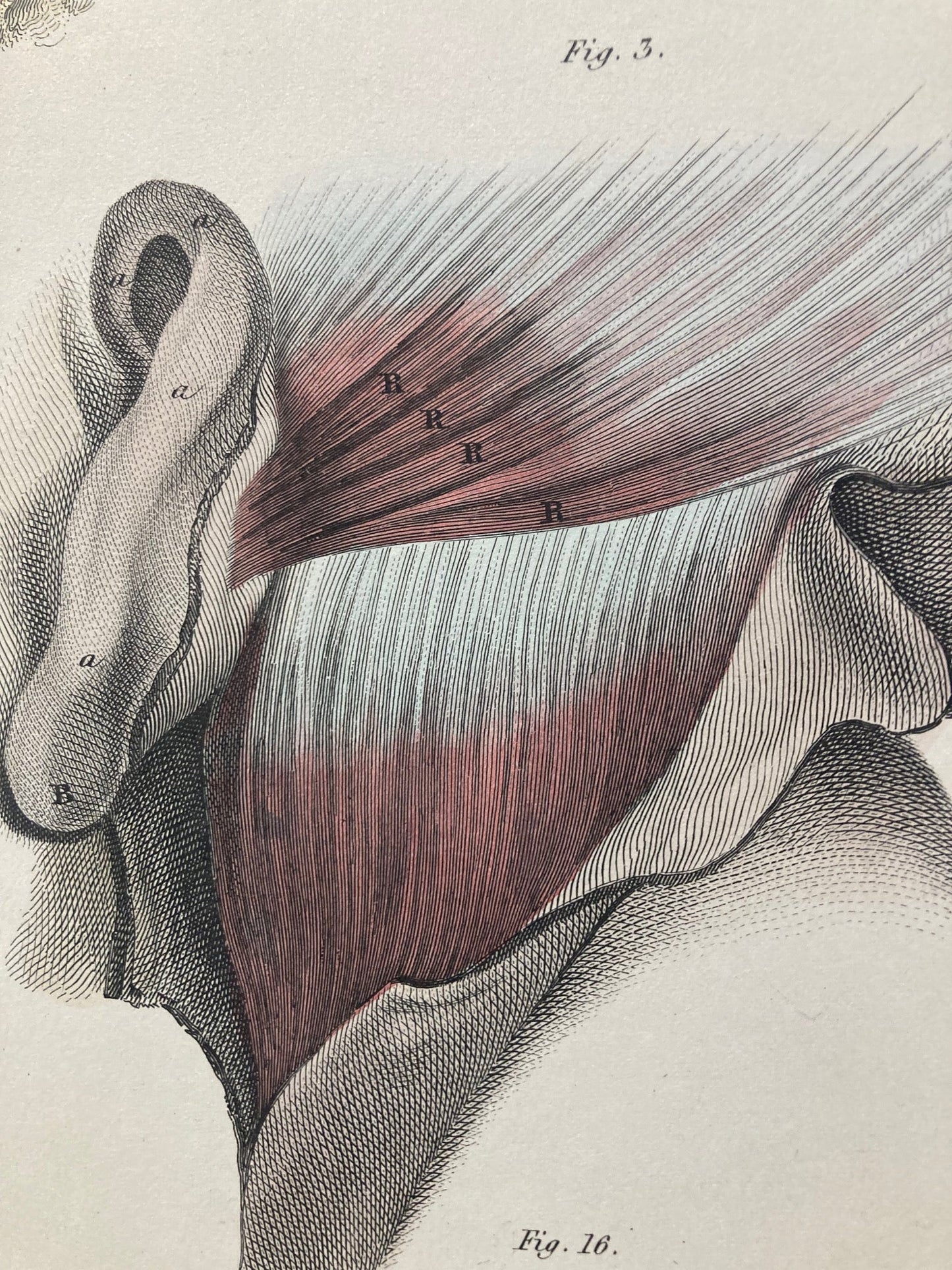
Originally published in a 19th-century medical atlas, this labeled plate highlights the auricle, ear canal, and surrounding tissues.
Visual Detail:
Includes labeled views of muscle fibers, nerves, and auditory structures—ideal for study or display.
Image & Format Notes:
Images shown are low-resolution previews. Full-resolution files or prints are provided upon purchase. Archival-quality materials used. Minor age-related wear may be present.
Use Cases:
Perfect for audiologists, educators, physiotherapists, or collectors of vintage medical art.
Condition & Format: Unframed.
Antique Good - Clean page, no foxing, bright detailed images- these plates have been around for almost 200 years. Minor age-related wear. Archival-quality paper. Dimensions: 44cm x 28cm
original 200 year old An hand-coloured drawing: [1823-1827] -John Lizars.
*John Lizars studied with Edinburgh surgeon John Bell and later taught anatomy and surgery in that city. John's brother William Home Lizars (1788-1859) engraved all of the plates on copper. "The book was costly to produce, for the engravings or etchings were, as usual, time-consuming, and the colouring was done skillfully and painstakingly by hand" (Roberts and Tomlinson, The Fabric of the Body, 504-505). "This superb atlas is certainly one of the most elegant works of the nineteenth century" (Cushing L313). Heirs of Hippocrates 1436; Waller 5950; Wellcome III: 531.
FREE Worldwide Shipping & Uplift Available
Share